
What is it?
Magnetic resonance imaging (MRI) is a type of technology used to take detailed pictures of the body. It is commonly used to detect abnormalities in the body, diagnose diseases, and to regularly monitor patients who are undergoing treatments. It can generate three-dimensional images of non-bony tissues, such as the brain. MRI procedures are non-invasive, require minimal preparation, and are not associated with health risks, as it does not use harmful types of radiation such as X-rays.
How does it work?
Human tissues contain water, which contain very small particles known as protons that behave like tiny magnets. An MRI machine uses large, powerful magnets to generate a magnetic field that can change how these particles rotate in your body, making them align with the magnetic field. Non-harmful radio waves are then pulsed through the patient, changing the direction of these particles, such that they are no longer aligned with the magnetic field. The radio waves are then turned off, and the particles can then re-align with the magnetic field. Different types of tissue and structures in the body will have particles that re-align differently, which can be detected by the machine to generate a detailed black and white image of the scanned area of the body. In addition to such structural information, MRI scans can provide information about how the brain is wired, levels of important chemicals, blood flow, metabolism, and brain function by acquiring information differently with the same machine.
How do you prepare for an MRI scan?
Since an MRI scan uses a large magnet, electronic devices and metal objects, such as glasses and jewelry, must be removed. There is usually no other preparation required for the scan. Patients must lie very still to generate a clear image. Patients do not need to be sedated, unless they have trouble lying still for the procedure. MRI scans that are obtained for research do not use anaesthesia to avoid unnecessary risk to research participants.
What happens during an MRI scan?
The patient lies down on a table that will move into the tunnel-shaped chamber. The patient is usually awake and will remain in the chamber as several scans are taken during the procedure (about 30-60 minutes). As the scan proceeds, there are often loud mechanical sounds, so earplugs are provided for protection. Some patients may experience claustrophobia, or are bothered by the noises. Becoming more familiar with the procedure, or listening to music or closing your eyes can help alleviate discomfort during the scan.
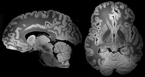
What do doctors look for in patients with SCAs?
MRI scans are often used to image the brain to detect signs of spinocerebellar ataxia (SCA), especially in a region of the brain known as the cerebellum. SCA is associated with brain cell loss, and appears as reduced volume of brain tissue in the MRI image.
If you would like to learn more about Magnetic Resonance Imaging (MRI), take a look at these resources by the National Institutes of Health and the Mayo Clinic.
Snapshot written by Dr. Claudia Hung and edited by Dr. Gülin Öz.

