Written by Dr. Terri M Driessen Edited by Dr. David Bushart
Mitochondrial dysfunction and loss of mitochondrial DNA is identified in an SCA1 mouse model.
Spinocerebellar ataxia type 1 (SCA1) is a neurodegenerative disorder that causes cell death in certain parts of the brain. The brain regions affected play important roles in motor coordination. The loss of coordination and movement – a symptom called ataxia – is the one of the primary effects of this disease. To investigate the causes of SCAs, researchers often use mouse models. In mouse models of SCA1, there are deficits in motor coordination before a significant amount of neurons (i.e., brain cells) are lost. This suggests that changes in neuron function, and not necessarily neuron death, may cause behavioral changes in SCA1. However, the mechanisms that cause dysfunction in SCA1 neurons are still a mystery.

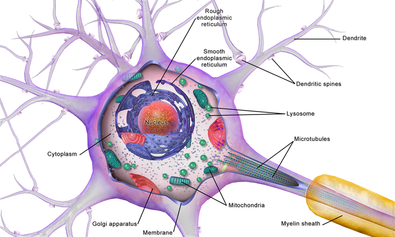
The brain requires a lot of energy to function. Without this energy, our neurons would be unable to survive. The cellular machines that generate this energy are the mitochondria, which are small organelles found in neurons (and nearly every other type of cell, for that matter). If the mitochondria in neurons do not function properly, this could lead to abnormal neuronal functioning. In fact, mitochondrial dysfunction has been found in several neurodegenerative diseases, such as Amyotrophic Lateral Sclerosis (ALS, or Lou Gehrig’s disease), Spinal Muscular Atrophy, Alzheimer’s Disease, Parkinson’s Disease, and Huntington’s Disease. Previous studies have also linked mitochondrial dysfunction to SCA1. It has been shown that Purkinje cells, the major cell type affected in SCA1, have altered levels of mitochondria-related RNA and proteins in SCA1 mouse models (Stucki, et al. 2016; Ferro, et al. 2017).
Mitochondria use a process called oxidative phosphorylation to make energy. In a recent study, Michela Ripolone and colleagues set out to determine whether changes in oxidative phosphorylation occur in SCA1. They found that Purkinje cells had reduced levels of oxidative phosphorylation components in early stages of disease. Interestingly, these findings are specific to Purkinje cells: another cell type called granule cells, which are located near Purkinje cells but have a different function, do not show a similar reduction in oxidative phosphorylation components.
Mitochondria use a specialized group of proteins called “complexes” that play a role in oxidative phosphorylation. Deficits in these complexes can cause mitochondrial dysfunction, which might lead to changes in the amount of energy produced by mitochondria. To determine if these complexes are affected in SCA1, Ripolone and colleagues examined their activity in SCA1 mice. They found significant changes in several of these complexes in SCA1 mice compared to control mice, suggesting that mitochondrial dysfunction might be important in SCA1.
Neurons contain DNA in the nucleus, but mitochondria also have their own DNA, which is called mtDNA. Since mtDNA provides a genetic code for mitochondrial protein complexes, Ripolone and colleagues wanted to determine if changes in mtDNA were present in SCA1 mice. They surveyed the levels of mtDNA in SCA1 Purkinje cells that had mitochondrial dysfunction, and Purkinje cells that appeared unaffected. They found that there was less mtDNA in the SCA1 Purkinje cells that had deficits in oxidative phosphorylation. These results were found in mice that were older. Importantly, in a separate study, it was found that no significant change in mtDNA occurs when these same SCA1 mice are younger (Sanchez, et al. 2016). This indicates that over the course of SCA1 disease progression, a decreasing amount of mtDNA in Purkinje cells might be an important feature of disease. However, the question remains as to what this reduction in mtDNA does to the cells. Ripolone and colleagues did not find a change in the number of mitochondria in SCA1 mice, indicating that the lower levels of mtDNA is not caused by fewer mitochondria overall. A previous study found that certain factors can impair mtDNA quality in SCA1, and even exacerbate symptoms of the disease (Ito, et al. 2015). With this prior evidence, and the work presented here by Ripolone and colleagues, it is plausible that mitochondria play an important role in SCA1.
Mitochondrial dysfunction has been found in a wide range of neurodegenerative diseases – not just SCA1 – which suggests that it could be a disease-causing factor in multiple different disorders. This means that, if mitochondria-focused treatments are developed, they might be useful for treating a variety of diseases. The results of Ripolone and colleagues have shed light on how mitochondria are affected in SCA1, contributing to a growing field of research of mitochondrial function in neurodegenerative disease.
Key Terms
Mitochondria: small organelles that are found inside of cells; produce the energy cells need to survive.
Oxidative phosphorylation: the main metabolic pathway that produces energy in cells. In humans, oxidative phosphorylation is carried out by mitochondria. Alterations in the oxidative phosphorylation system can lead to a reduction in the amount of energy available to a cell, which can affect cellular function.
Purkinje Cells: one of the primary cell types affected in SCA1. These cells are located in the cerebellum, where they play a role in regulating motor coordination.
Conflict of Interest Statement
Dr. Terri M Driessen has no conflicts of interests to declare. Dr. David Bushart has no conflicts of interest to declare.
Citation of Article Reviewed
Ripolone M, et al. Purkinje cell COX deficiency and mtDNA depletion in an animal model of spinocerebellar ataxia type 1. J. Neurosci. Res. 2018 96(9):1576-1585. https://www.ncbi.nlm.nih.gov/pubmed/30113722


