
Written by Dr. Ambika Tewari Edited by Dr. Sriram Jayabal
Targeting phosphatases in the cerebellum can correct miscommunication in multiple models of ataxia.
The cerebellum is essential for motor coordination and consists of the coordinated activity of different types of cells. Purkinje cells are one of the most fascinating cell types in the cerebellum. They have an elaborate network of branches called dendrites, where a neuron receives communication from other neurons. It is one of the most complex branching systems seen across all neurons in the entire brain. Each one of these branches has many points of contact with other branches called axons. Each axon is part of a neuronal structure that allow communication between neurons. These axons are from different cell types and allow information to be transferred to Purkinje cells.

Due to this branching complexity, Purkinje cells receive many messages or inputs. This represents different pieces of sensory information to ensure that movements are precisely timed. Purkinje cells must integrate and process this information. This produces motor behaviors like walking, writing, playing a musical instrument, and many more. Any alteration to the processing of this information will result in cerebellum dysfunction; in fact, Purkinje cells have gained attention because they undergo progressive deterioration in most ataxias.
Neurons, including Purkinje cells, communicate with other neurons using electrical signals known as action potentials or spikes. Firing rate, defined as the number of spikes within a defined period of time, is thought to be an important feature of this communication, which is critical for coordinating muscle movements. Therefore, a lower firing rate in Purkinje cells would signal a faulty communication between Purkinje cells and their targets. This has devastating consequences as seen in many ataxias.
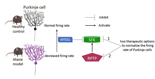
For instance, in an earlier study, a group of authors found that the firing rate of Purkinje cells was decreased in mouse models of three different Spinocerebellar ataxias (SCAs): SCA1, SCA2, and SCA5. They further explored whether there was a common reason underlying the decreased firing rate. They found that a protein named Missing in Metastasis (MTSS1), was important for Purkinje cells to effectively communicate with each other. Mice engineered to have no MTSS1 protein had a decreased firing rate and difficulty walking and maintaining their balance.
In every cell in the body, including brain cells, there are numerous proteins that perform different functions. The concerted effort of all are needed for the cell to perform its intended duty. Some of these proteins are maintained in the cell in an inactive form and are activated when they are required in the cell and inhibited when they are not. This highly regulated system aims to maintain precise levels of proteins in each cell, while simultaneously conserving energy. Each cell has many ways of activating/inactivating a protein. A specialized group of proteins known as kinases and phosphatases, adds and removes phosphate groups to and from proteins respectively, thereby altering their active/inactive forms which then changes their interactions with other proteins. MTSS1 is one such protein that inhibits the activity of a group of kinases known as Src family of non-receptor tyrosine kinases (SFKs).
In the study mentioned earlier, elevated SFK levels were found in the cerebellum of SCA1, SCA2, and SCA5 mouse models. When researchers treated these ataxic mice with an inhibitor of SFK, they found that their walking ability was improved. This inhibitor, Dasatinib, while FDA approved, does not cross the blood-brain barrier, which creates a protective shield around the brain. In mice, the drug was delivered directly to the brain and continuously infused to maintain its levels. In human subjects, however, this is a highly risky procedure that is not recommended because of its invasiveness. Therefore, the same group of researchers embarked on a quest to find another way to inhibit SFKs owing to its therapeutic potential in MTSS1 mutant mice. The researchers found a group of proteins known as Receptor-Protein Tyrosine Phosphatases (RPTPs), which lead to the activation of SFK.
To test their hypothesis that inhibiting RPTP would in turn prevent the activation of SFK, the researchers performed a series of experiments using cells cultured in a dish. By adding a particular combination of nutrients to the cells, these cells can continue to live, grow and divide when stored at the right temperature. When the cells were treated with an inhibitor against RPTP, the researchers observed a decrease in SFK activity, supporting their hypothesis.
They next wanted to test the inhibitor’s effect on the firing rate of Purkinje cells in mouse models with ataxia. The researchers used two models from their earlier study: one that had no MTSS1 protein and a mouse model of SCA2. They prepared slices from the cerebella of these animals and recorded the firing rates of the Purkinje cells, while the slices were bathed in a solution that contained the RPTP inhibitor. Inhibiting RPTP rescued the firing rates in both mouse models to levels comparable to the control mice that had no obvious Purkinje cell firing deficits. These results show that multiple ways could be implemented to maintain healthy levels of SFK to ensure proper communication between Purkinje cells and its target.
In summary, the authors successfully uncovered an alternative method to normalize the firing rate of Purkinje cells in these mutant mouse models. By targeting another group of proteins, the RPTPs, which activate SFK, they could reproduce the data that they observed when SFK was directly inhibited. One crucial question that remains unanswered is whether the mice treated with this inhibitor will improve in walking ability. Further, future research is needed to test this drug, and to find other inhibitors of RPTPs, to assess their efficacy as a potential therapy in long-term studies.
Key Terms
Action Potential: The electrical signal that nerve cells use to communicate with each other
Kinases: A specialized type of protein that adds phosphate groups to proteins, altering its interactions with other proteins
Phosphatases: A specialized type of protein that removes phosphate groups from proteins, altering its interactions with other proteins
Inhibitor: a substance that slows down or prevents the normal or default reaction from taking place
Blood-Brain Barrier: A physical barrier that separates the bloodstream from the brain creating a highly restrictive area that prevents entry of pathogens into the brain
Invasiveness: The entry of a foreign object into the body by incision or puncture that require trained healthcare professionals
Conflict of Interest Statement
The author and editor declare no conflict of interest.
Citation of Articles Reviewed
Brown, A.S., et al. Receptor protein tyrosine phosphatases control Purkinje neuron firing. Cell Cycle, 2020. 19:2 (https://doi.org/10.1080/15384101.2019.1695995)
Brown, A.S., et al. MTSS1/Src family kinase dysregulation underlies multiple inherited ataxias. Proceedings of the National Academy of Sciences, 2018. https://doi.org/10.1073/pnas.1816177115

