Written by Katerina Pliatsika
Edited by Hannah K Shorrock, PhD
Additional proteins which interact with ATXN1 contribute to toxicity in a SCA1 mouse model.
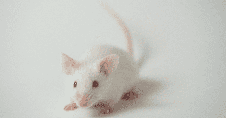
In neurodegenerative diseases, a problematic gene and its resulting protein play a crucial role in the selective loss of brain cells. However, a fundamental question remains unanswered: even though the faulty protein is present throughout the brain, why are only certain neurons are affected? Dr. Zoghbi and her research team tried to answer this question by studying a mouse model of Spinocerebellar ataxia type 1 (SCA1). Their findings challenged the common belief that SCA1 mainly impacts the cerebellum, revealing that other brain regions are also affected by the expanded polyQ ATXN1. Importantly, they uncovered new proteins that are involved in the progression of this disease.
SCA1 affects approximately one in 100,000 individuals worldwide and is characterized by imbalance and movement disabilities. It is caused by a repeat expansion of three DNA bases, C, A and G, in the ATXN1 gene. In the protein produced from this gene, also called ATXN1, the CAG repeat results in multiple units of the amino acid glutamine next to each other which is called a polyglutamine, or polyQ, tract. This abnormal expansion leads to neuronal degeneration mainly in the Purkinje cells of the cerebellum. We know that without the polyQ tract, ATXN1 normally interacts with a protein called Capicua (CIC). Capicua is a transcription factor which means it is responsible for regulating the expression of many genes. However, previous studies show that the polyQ tract in the SCA1 ATXN1 protein disrupts the normal relationship between Capicua and ATXN1 leading to too much reduction in the levels of capicua-regulated genes. This excess reduction in expression ultimately proves lethal to neurons. Importantly, by disrupting the interaction between capicua and the SCA1 ATXN1 protein, the research team previously showed that they could correct the harmful effects caused by this interaction in SCA1 mouse cerebellum.
Combining the information the research group had learned from their previous study and two key pieces of information,
1) ATXN1 is widely expressed throughout the brain; and
2) Brain regions beyond the cerebellum, including the brainstem, are affected in SCA1 and degenerate as the disease progresses, the group identified a new research question: Is the interaction between capicua and SCA1 ATXN1 protein also responsible for the loss of brain cells and disruption seen in these other brain regions?
To address this question, the research group generated a new mouse model. They took a mouse model that produces a mutant ATXN1 with an extended sequence of 154 glutamines and mutated two critical amino acids. These amino acids were located at the position where ATXN1 interacts with capicua. In making these mutations, the researchers prevented the interaction of these two proteins throughout the brain. This approach allowed them to assess whether the interaction, crucial for degeneration in the cerebellum, also impacted other brain regions.
The researchers looked at whether the disruption of SCA1 ATXN1 capicua interaction led to improvements in SCA1 neurological phenotypes across the entire brain in this mouse model. In the cerebellum, the researchers found improved neuronal organization, connectivity and function after mutation of the two key amino acids. Next, they examined whether loss of the ATXN1-capicua complex could alleviate other SCA1-related symptoms, such as kyphosis and weight loss. They found that these symptoms were not improved. Similarly, learning and memory deficits persisted despite the loss of ATXN1-capicua complex. However, the researchers found a partial improvement in other symptoms, including motor incoordination, respiration and lifespan. While these findings demonstrate that the SCA1 ATXN1 capicua interaction is important for some phenotypes, this research also suggests the involvement of additional factors in the progression of the disease.
In order to understand what these other factors might be the researchers examined the impact of the amino acid mutations on changes in gene expression across the brain in SCA1. Because capicua is a transcription factor responsible for turning on and off many genes, using RNA-Sequencing could show which genes were rescued by the amino acid mutations that disrupt the interaction between ATXN1 and capicua and which genes are not rescued. The research group looked at this in five different regions: the cerebellum, hippocampus, brainstem, cortex, and striatum. They observed changes in the level of genes in all brain regions when comparing SCA1 mice with wild type mice. While disruption of the ATXN1-capicua complex improved a variety of events across all tissues in SCA1 mice, it didn’t fully normalize all changes in gene levels. These findings suggest that while other studies have demonstrated that the ATXN1-capicua interaction is important in mediating dysfunction in the cerebellum, there are also other factors contributing to the disease brain-wide.
The findings that the disruption to the ATXN1-capicua complex only partially improved gene activity changes prompted the researchers to investigate the involvement of other key proteins interacting with ATXN1. They hypothesized that these proteins might play a role in SCA1 toxicity in other brain areas, beyond the cerebellum. To answer this question, researchers used a mouse model that produces ATXN1 with two glutamines and carries mutations in the two amino acids that are critical for its interaction with protein capicua. To investigate whether other proteins besides capicua interact with ATXN1, they used a technique that allowed them to isolate ATXN1 along with any proteins attached to it. Through this they discovered three brain-expressed novel transcriptional factors – zinc finger with KRAB and SCAN domains 1 (ZKSCAN1), zinc finger and BTB domain containing 5 (ZBTB5) and regulatory factor X 1 (RFX1), that interact with both wild-type and polyQ expanded ATXN1 even in the absence of capicua binding. The researchers found that two of these, RFX1 or ZKSCAN1, control many genes that are abnormally turned on or off in the SCA1 mouse model. Even when the interaction between ATXN1 and capicua is blocked, many genes stay abnormal likely because RFX1 and ZKSCAN1 remain bound to ATXN1. Importantly, the researchers found that genes regulated by these transcription factors in the mouse model also showed similar abnormalities in neurons derived from SCA1 patients, suggesting these findings are relevant to human disease. Together with capicua, these newly discovered proteins are predicted to regulate approximately 33% of the affected genes in SCA1.
In conclusion, this work has significantly advanced our understanding of the mechanisms driving the progression of SCA1. What is interesting about this study is that it doesn’t focus only on the cerebellum, which is traditionally linked with SCA1, but it expands our interest into other brain regions. The discovery of additional proteins that interact with ATXN1 holds promise for the development of targeted interventions and treatments for this disorder. By understanding the diverse factors driving SCA1 pathology brain-wide, we move closer to a future where effective therapies offer hope for individuals and families affected by this neurological condition.
Key Words
ATXN1: A protein which is encoded by the ATXN1 gene. Mutations in the ATXN1 gene are associated with the neurodegenerative disease SCA1.
Cerebellum: Located at the back of our head, the cerebellum is responsible for coordinating movement and maintaining balance. It is the most affected brain area in Spinocerebellar ataxia type 1.
ATXN1-CIC complex: The interaction of the mutant ATXN1 with the Capicua (CIC) protein drives toxicity in cerebellar cells.
Conflict of Interest Statement
The author and editor have no conflicts of interest to declare.
Citation of Article Reviewed
Coffin, Stephanie L et al. “Disruption of the ATXN1-CIC complex reveals the role of additional nuclear ATXN1 interactors in spinocerebellar ataxia type 1.” Neuron vol. 111,4 (2023): 481-492.e8. doi:10.1016/j.neuron.2022.11.016
Read Other SCAsource Summary Articles

RFC1 Ataxia (CANVAS): Modifiers and predictors of disease onset and severity
Written by Tala Ortiz Edited by Hannah Shorrock, PhD Repeat length in RFC1 Ataxia, also known as CANVAS, impacts the age of onset of symptoms, type of symptoms, and progression Are there ways to Read More…

Unraveling the Role of Mitochondria in SCA6 Progression
Written by Yujia Li Edited by Priscila Pereira Sena The discovery of first links between mitochondrial dysfunction and SCA6 progression offers new insights for potential treatments. What drives the progression Read More…

Treating Disease Mutations at the Source: A New Paradigm of Treatment for Spinocerebellar Ataxias
Written by Ross Pelzel Edited by Eder Xhako, PhD and Celeste Suart, PhD Alternative splicing produces different versions of proteins that arise from the same gene. In many neurodegenerative diseases, Read More…


