


Skip to content

Join the Summer Match Challenge! All donations during the month of June will be matched by the Clementz-Peterson Family Fund and an anonymous donor, up to $100K. DONATE

Snapshot: What is a CT Scan?

A computer tomography (CT) scan, also called a CAT scan, is a diagnostic imaging procedure that uses X-rays and a computer to help doctors see inside your body.  CT scans provide detailed, cross-sectional pictures of bones, soft tissues, and organs that doctors can use to detect problems like injuries, infections, or diseases.

CT scans generate more detailed images than standard X-rays, where some features of internal organs and other structures are not visible. In a standard X-ray, a single beam of energy is aimed at the tissue that is being studied. A CT scan uses a large machine that rotates around your body taking many x-ray images from different angles. A computer then combines these images to create detailed two-dimensional “slices” of the body. Doctors can look at each slice individually or stack them together to form a clear, three-dimensional view of what is happening inside. 

By capturing different views of an organ or other tissue, a CT scan provides detailed information about it. This information can help doctors detect tissue abnormalities, internal bleeding, injuries, tumors or infections. CT scans are also often used to guide medical procedures like biopsies and to monitor health conditions over time.

CT scans sometimes can be done using contrast. Contrast is a special dye, taken my mouth or injected into an IV, that helps doctors visualize certain tissues more clearly.

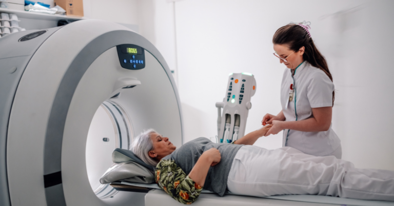
A CT scan is quick, non-invasive, and painless. You will need to lie still and may be asked to hold your breath for a few seconds. The large, doughnut-shaped X-ray tube may make soft whirring sounds as it moves around you, but it does not touch you. CT scans use small amounts of ionizing radiation, but the levels used are considered safe and kept as low as possible. The valuable information gained from these scans often helps doctors detect and treat conditions more accurately, leading to better care and outcomes.

CT scans and Spinocerebellar Ataxia

Although a CT scan alone cannot be used to definitively diagnose spinocerebellar ataxia (SCA), doctors will often use a CT scan to rule out other causes of ataxia in a new patient, such as a mass or bleed in the brain. A doctor may also use CT scans to check for structural changes in the cerebellum or other parts of the brain that control movement and balance. CT scans are also valuable in research settings, where scientists may use them to monitor the impact of treatments.

If you would like to learn more about CT Scans, take a look at these resources by Johns Hopkins, Mayo Clinic, or Cleveland Clinic

Snapshot Written by:Asmer Aliyeva

Edited by: Chloe Soutar, PhD.

Read Other SCAsource Snapshot Articles

Snapshot: What Are Silent Mutations?

Our DNA (deoxyribonucleic acid) serves as a genetic blueprint for building and maintaining our tissues. This complex molecule contains the information needed to build proteins, written in a code made Read More…

Translate »

Join the Ataxia community today!

Become a free member for exclusive content from NAF.