
What are C. elegans?
If you read the title of this article and had no idea what Caenorhabditis elegans are, you are not alone! Caenorhabditis elegans, more commonly known as C. elegans, are microscopic worms that typically grow up to 1 mm in length. C. elegans are naturally found worldwide in soil where there is rotting vegetation. If you are feeling brave, you can try to locate them in your household compost! Although these worms are less familiar to the general public, C. elegans are well known to scientists, since studying these tiny worms has taught us a lot about human disease.
Why are C. elegans used as a model system?
C. elegans were first isolated in 1900 and, since the late 1960s, have been used to “model” human disease. This is because C. elegans and humans share some common physiological features and have a significant overlap in their genetic codes. SCAsource previously published a Snapshot on mouse models, which are widely used in ataxia research,. Although C. elegans are not used as widely in ataxia research, there are many advantages to using C. elegans as a model system:
- C. elegans are inexpensive to maintain, allowing for the screening of thousands of drugs at a relatively low cost. Once administered, scientists can study the drugs’ effects on C. elegans movement, development, and nervous system function.
- C. elegans are easy to grow in the laboratory.
- C. elegans are self-fertilizing hermaphrodites, meaning that they can reproduce without a sexual partner. A single hermaphrodite can produce 300-350 offspring over a 3-day period, allowing scientists to easily study a large number of worms that have the same genetic characteristics.
- Scientists can easily manipulate the genome of C. elegans to study many human diseases.
- Because C. elegans are transparent, their internal organs, including the nervous system, can be imaged without dissection.
How can C. elegans be used to study neurodegeneration?
The nervous system of a C. elegans is made up of a few hundred neurons, which is relatively simple compared to the human brain (which contains about 86 billion neurons). Because of this simplicity, scientists have used C. elegans to develop models for several neurodegenerative diseases, including Alzheimer’s, Parkinson’s, Friedreich’s ataxia and, more recently, spinocerebellar ataxia type III (SCA3). The SCA3 C. elegans model was developed by a research group in Portugal led by Dr. Patrícia Maciel, and it is the first of its kind in the spinocerebellar ataxia field. These worms express the human SCA3-causing protein in all their neurons, resulting in adult-onset motor dysfunction that resembles what we see in SCA3 patients.
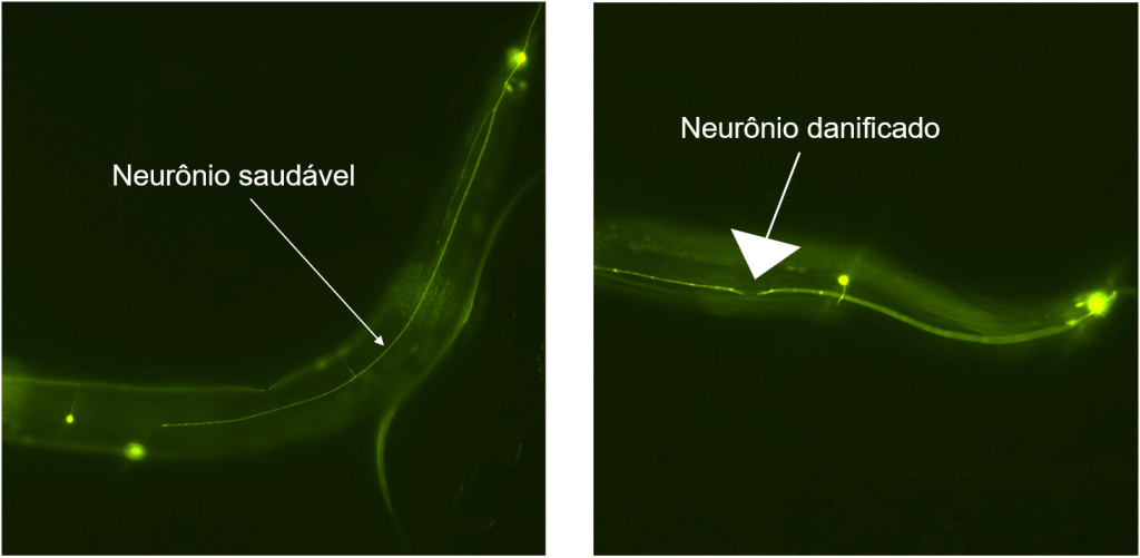
Neurodegeneration (damage/death of neurons) in C. elegans is monitored by tagging neurons with a marker that shines green under a specific type of light. The health of neurons is then assessed, making it possible to determine if neurodegeneration has occurred. The image above shows a healthy C. elegans neuron on the left, which appears intact, compared to a damaged C. elegans neuron on the right, which has a break (white arrowhead). Being able to distinguish between healthy and damaged neurons in C. elegans is very useful, as scientists can use this tool to test different ways of repairing or protecting neurons. If scientists are able to slow or prevent neurodegeneration in C. elegans, there is potential that such a discovery could eventually help treat human neurodegeneration, as well.
I hope this short summary has shown you that there is a massive amount of scientific potential in these tiny worms! Understanding the biology of C. elegans provides insight into human biology, like how neurodegeneration occurs and what we can do to stop it.
If you would like to learn more about C. elegans model systems, take a look at WormBook, Wormbase, and WormAtlas.
Thank you to Kim Pho from Dr. Lesley MacNeil’s lab at McMaster University for providing the fluorescent images of C. elegans neurons.
Snapshot written by Katie Graham and edited by Dr. Lesley MacNeil.

